Back Pain Imaging and Exercise
Spinal imaging shows us much about a Richmond back pain and leg pain sufferers’ condition. Imaging must be reviewed carefully, of course, for what it reveals as well as the way various imaging methods compare to others. Johnson Chiropractic appreciates all the input from you, the Richmond back pain patient, as well as your imaging and exam findings to individualize your Richmond chiropractic treatment plan for pain relief.
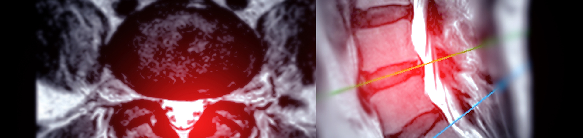
DISC HERNIATION AND Richmond BACK PAIN
Disc herniation and leg and/or back pain seem to come together, too, but so much more may be involved: disc degeneration, ruptured annulus fibrosus, irritated and compressed nerve roots and cauda equina from the herniation itself. A newer study tracked the effects of exercise on a disc herniation’s neuromechanical compression, its inflammatory chemical stimulation, and its autoimmune response. (1) Imaging demonstrates nice pictures of the disc herniation…that Johnson Chiropractic correlates with your symptomatology!
BACK PAIN AND IMAGING
Back pain and imaging tend to go together. When someone experiences back pain, xrays, MRIs, and CTs come to mind. Just what do these display? A study compared images of back pain patients to asymptomatic persons. MR imaging of those with back pain showed that a variety of measurements differed: anteroposterior diameter dimensions of the vertebral canal varied, transverse diameter was lesser, and thecal sac area was smaller. (2) Caution is necessary to compare neuroforaminal measurements from CT scans and plain film as a study found that in patients without back pain plain film measurements were bigger compared to those on CT. (3) Another imaging finding your Richmond chiropractor is quite aware of is called Modic change. It’s another imaging finding that is very revealing in that Modic change (mainly type II) is related with abdominal aortic calcification via a lower blood supply or even poorer systemic vascularization because of atherosclerotic disease. (4) It’s another insight we can utilize to determine what’s going on in your spine. Finally, a spinal imaging finding that is quite usual in back pain patients is fatty infiltration of the paraspinal muscles, more so in patients with degenerative lumbar spondylolisthesis. (5) We check the MRI images for that though it’s hard to not see! Another good reason to start a good spinal exercise program!
EXERCISE & SPINE PAIN RELIEF
Exercise may be quite effective in managing the lumbar disc herniation which is why Johnson Chiropractic incorporates some simple lumbar spine exercises to begin on day one. We inspire our back pain patients to continue performing these simple exercises and are happy to support anyone who wants to do more...after we talk!
CONTACT Johnson Chiropractic
Listen to this PODCAST with Dr. Lee Hazen on The Back Doctors Podcast with Dr. Michael Johnson as he describes a case of degenerative spondylolisthesis that responded well to treatment with The Cox® Technic System of Spinal Pain Management.
Schedule your Richmond chiropractic appointment today to see us! Whether you have spinal imaging already or need it, your Richmond chiropractor can help make sense of its significance in your journey to back pain relief.